
Scientific analysis on boards
Révéler l’invisible d’un tableau à l’aide de l’imagerie scientifique. L’analyse scientifique des tableaux est indispensable pour dater le support, observer une signature cachée, un repentir ou un dessin préparatoire, et identifier les pigments. Découvrez les différentes méthodes d’imageries utilisées par les laboratoires CIRAM afin de révéler l’invisible de vos tableaux.
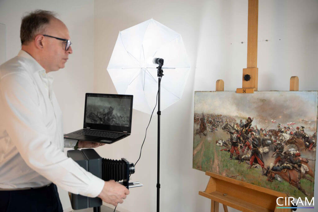
Whether for restoration, conservation or authentication, scientific analysis of paintings is an important and unavoidable step.
First and foremost, the different materials of a painting must be considered to deliver a reliable interpretation. Our scientists date the support (canvas, wood, paper, cardboard…), analyze pigments, mineral fillers, binders and varnishes. To achieve this, CIRAM laboratories carry out a complete study of paintings using carbon-14 dating, chemical analysis and scientific imaging techniques.
The different scientific imaging techniques
Scientific imaging techniques provide invaluable information about a painting. They enable the investigation of all the layers of a painting, from the varnish, to the support, via the preparatory drawing, pictorial layers, repentirs, repaints and any restorations. Natural light, ultraviolet, infrared and X-ray radiography, discover the full range of imaging techniques for analyzing paintings.
Visible light imaging
We distinguish three different kinds of visible light examination:
- Visible light photography: produced under specific conditions, these images correspond to classic photography;
- Grazing light photography: this type of photography reveals the state of conservation of the work’s surface. Our scientists illuminate the work with a focused beam of light forming a 15° angle with its surface. This technique enables us to identify alterations to the paint layer (lifting, blistering, cracking, etc.), deformations of the support (poor tension of the canvas, cracks or joining of the boards) and alterations such as tears, scratches or dents. Grazing light also provides clues to painting techniques and the painter’s style.
- Transmitted light photography: this is observed on the reverse side of canvas or paper paintings. With this method, our scientists apprehend any holes and tears, it also reveals alterations to the pictorial layers.
Ultraviolet (UV) radiation)
Ultraviolet (UV) radiation lies outside the visible spectrum, and is divided into near UV (380nm to 200nm) and extreme UV (200nm to 10nm).
UV imaging enables the surface layer of the painting to be explored. It uses the fluorescence properties of the varnish, which is more or less intense depending on its composition and alteration. Overpainting on varnish or restorations can be easily spotted.
Important: in the absence of varnish, certain natural pigments can emit a colored fluorescence; this fact must be taken into account to avoid distorting interpretation.
Infrared radiation (IR)
Infrared radiation is located between 780nm and 5mm, beyond the visible spectrum, but on the other side of the ultraviolet. CIRAM laboratories use a so-called warm” light emission that lies in the near infrared. This makes it possible to record the absorption of infrared radiation by the materials making up the pictorial layers.