

OUR IMAGING SERVICES
TECHNIQUES ADAPTED TO YOUR ART AND HERITAGE OBJECTS
We use different scientific imaging techniques, natural light, grazing light, transmitted light, UV fluorescence, infrared reflectography, X-ray radiography and CT scan, to respond to your multiple problems.
These numerous techniques allow us to visualize the invisible: a hidden signature, the contents of a cinerary urn, a preparatory drawing, a restoration area, a repentance, an old painting…
Do you want to analyze your products?
Our experts will help you choose the techniques to use and answer your questions
Request a studyPrecision imaging technologies
CRIAM laboratories offer scientific imaging techniques that can be used to study paintings, wall paintings, drawings, but also stone or metal sculptures, and also archaeological furniture made of wood, terracotta, metal.
CRIAM laboratories accompany you in your problems whether they are associated with the art market, heritage conservation or archaeology. For more information, request a quote via our form.
Scientific imaging for your art objects
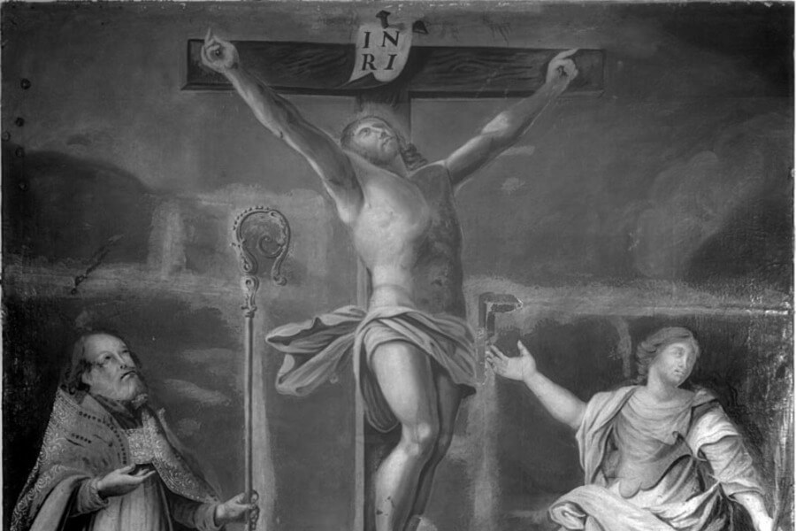
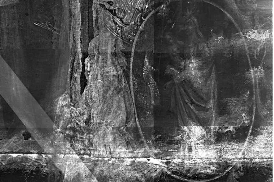
The infrared reflectography, UV fluorescence or X-ray radiography are complementary techniques for studying your painted works in general, and your paintings in particular. They will allow an in-depth analysis (from the surface varnish to the canvas or wood support) and will be indispensable investigations for the objective analysis of your paintings and drawings.
Whether it be restorations, a preparatory drawing, repentis or even a hidden signature, scientific imaging will reveal the invisible in your pictorial works.
Valuable analyses in archaeology
Thanks to advances in science, it is now possible to transport certain devices on site. CIRAM laboratories have portable devices allowing analysis directly on the archaeological site. This technical revolution concerns X-ray radiography, as well as infrared reflectography or UV fluorescence.
Associated with these portable techniques, CT scan or computed tomography is the essential tool for scientific imaging in archaeometry. It will reveal the contents of a cinerary urn or the internal structure of a gangue for example. Finally, CT scan allows virtual excavations.
X-ray radiography
The X-rays will pass through the material and be more or less absorbed depending on the density of the materials. This in-depth investigation of the objects reveals their homogeneity, their internal structure, the possible presence of exogenous materials, cracks or breaks, the different layers of a painting.
The devices made available by CIRAM are either portable, thus usable in situ, or laboratory. This makes it possible to respond to all your problems of authentication, enhancement and conservation of heritage.
Infrared imaging, a gold mine for paintings
Infrared reflectography is an indispensable part of analyzing your pictorial works. This HD and portable digital camera is used to:
- Detect areas of restoration;
- Identify landmarks to understand the stages of creation of the painting or pictorial work;
- Visualize preparatory drawings;
- Detect signatures or false signatures with a view to authentication.
Infrared reflectography will highlight elements invisible to the naked eye and essential to the exhaustive study of an easel painting or a mural.
CT scan and Computed tomography
In addition to X-ray radiography, CIRAM’s scientific teams offer 3D studies by CT scan. They will allow to visualize the internal structure of heritage objects and to identify the content of archaeological furniture.
For the art market, CT scan will be used in addition to thermoluminescence to ensure that terracotta objects have not been reconstructed, or to identify the elements present inside a fetish for example.
For archaeology, CT scanning, in addition to identifying areas of fragility and the degree of conservation of objects, will allow virtual excavations.
UV fluorescence
Ultraviolet (UV) radiation lies outside the visible radiation spectrum and is divided into near UV (380 nm to 200 nm) and extreme UV (200 nm to 10 nm). UV imaging is an analytical technique that allows the exploration of the surface layer of a painting.
It uses the fluorescence properties of the varnish more or less intense depending on its composition and alteration (oxidation).
On a homogeneous background, an area of different intensity implies a variation in the nature of the varnish, potential evidence of a cleaning or restoration. In the absence of varnish, some natural pigments will be able to emit a colored fluorescence.